
Diagnostic Imaging - Veterinary Referral Centre
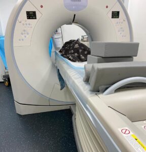
Computed Tomography (CT)
Indications for CT
- Lung masses or metastasis
- Interstitial lung disease e.g. fibrosis
- Mediastinal masses or effusion
- Unexplained pleural effusions
- Idiopathic pneumothorax
- Nasal discharge/epistaxis
- Orbit: Retrobulbar masses, foreign bodies
- Ectopic ureter
- Occult lameness – elbow
- Complex skeletal lesions e.g. vertebral, pelvic,
tempormandibular or skull fractures - Bone tumours
- Portosystemic shunts
- Search for occult neoplasia
X-rays are present throughout the universe passing through space but are only present in very low quantities surrounding us as we go about our daily duties on the surface of planet Earth. However, we knew nothing about them until Wilhelm Roentgen discovered their existence in a laboratory in Germany in 1895. When he discovered them however, he very quickly recognised their huge potential in the field of medicine. X-rays would for the first time in the history of the human race, allow mankind to look inside the human body and indeed animal bodies without having to cut them open! It was this discovery that resulted in Roentgen being awarded the first Nobel Prize for Physics in 1901.
Computed tomography or CT uses the same x-rays that Roentgen discovered in the 1890s but in a different way to produce a different type of image. The ability to use x-rays to produce a CT image was only made possible by the advent of another tremendous invention – the computer. Many people nowadays are familiar with CT having seen it on television or indeed having had a CT scan themselves. The patient lies flat on a table in the centre of a tunnel. So what happens now?
Imagine yourself for a moment at the centre of a large Ferris wheel like the London Eye for example. Except this Ferris wheel has 360 gondolas… You are lying still on your back looking up at the sky and all around you – above, below and to either side – are all these gondolas, each packed with sharphooters armed with magic x-ray guns. Once the operator presses the button to start the wheel, all the sharpshooters start firing at you at once and continue doing so as the wheel rotates around you. Luckily, you don’t feel anything because the x-rays pass through you magically. Phew! OK, what happens next?
Because x-rays are being fired from 360° around the patient instead of a single static point, things are far more complicated than with a regular x-ray examination. The computer ‘sees’ the x-rays passing through the patient from all these different directions and is then able to construct a picture of a slice of the patient just as you would slice through an orange. This is very different to a regular x-ray which is more like a photograph of the orange for example. This provides a major advantage over regular x-rays as the image avoids the superimposition of tissues which is inevitable on a regular x-ray. Because the CT scan produces an image of a slice or cross-section through the patient, it is often referred to as cross-sectional imaging. Magnetic resonance imaging or MRI is also a form of cross-sectional imaging because it to produces images of slices through the body.
A CT image differs from a regular x-ray image in another fundamental way. A regular x-ray shows fluid and all types of soft tissue (blood, urine, muscle, liver, spleen) as the same colour or opacity on the x-ray film. A CT image however is able to differentiate, not only between fluid and soft tissue, but different types of soft tissue. Thus, blood in the heart can be differentiated from the heart wall, tumour is from the surrounding liver, bleeding or blood clots in the brain and so on.
In veterinary medicine, CT scans are becoming more available to patients and vets and are being used more commonly as the primary method to diagnose orthopoedic diseases such as poorly-mineralised or fragmented medial coronoid process, distal humeral OCD, elbow incongruency and incomplete ossification of the humeral condyles (IOHC) or in the search for spread of cancer to the lungs. Complex anatomic areas such as the skull or spine are frequently evaluated in more detail with CT as it avoids the problem of superimposition seen with regular x-rays. The chest and abdomen are also evaluated more frequently when more detail is required subsequent to an abnormality being seen initially on a regular x-ray or ultrasound examination.
X-Ray
X-Rays are commonly used in veterinary medicine to investigate disease. X-rays are a type of energy radiating in waves throughout space and are a special form of radiaton called electromagnetic radiation. Other forms of electromagnetic radiation include radiowaves, microwaves, infrared light, visible light, ultraviolet light and gamma rays.
X-rays are present throughout the universe passing through space but are only present in very low quantities surrounding us as we go about our daily duties on the surface of planet Earth. However, we knew nothing about them until Wilhelm Roentgen discovered their existence in a laboratory in Germany in 1895. When he discovered them however, he very quickly recognised their huge potential in the field of medicine. X-rays would for the first time in the history of the human race, allow mankind to look inside the human body and indeed animal bodies without having to cut them open! It was this discovery that resulted in Roentgen being awarded the first Nobel Prize for Physics in 1901.
Care has to be taken when x-rays are used. Many people are familiar with the use of lead gowns when an x-ray is being taken for example. This is to protect parts of the body that are not being imaged from being exposed to the x-rays themselves. The risk is miniscule because the dose of x-rays from an x-ray procedure in a hospital or verterinary surgery is tiny. But there is no point is taking risks!
The reason that x-rays are potentially harmful is that they have the ability to ionise cells or atoms that they pass through. This means that they have enough energy to knock an electron out of the atom or cell (a bit like a billard ball being hit by the white ball) thereby damaging or killing it. This is only of significant risk if the dose of x-rays is high. Indeed this harmful property is put to very good use in both human and veterinary medicine in the form of radiation therapy. This is where x-rays and sometimes gamma rays, are focused carfully on a particular area and used to kill tumour cell so helping to treat and potentially cure cancer.
In veterinary medicine, x-rays are used commonly as the first step in diagnosing disease of bone such as arthritis, trauma, cancer, congenital and developmental abnormalities and metabolic bone disease. As far as nasal, orbital, spine, skull and brain disease is concerned, CT and MRI are more and more commonly being used as the primary investigative tool.
Examples of such bone disease include arthritis or degenerative joint disease, osteomyelitis (inflammation or infection of the bone and bone marrow), fractures, or dislocations, osteosarcoma, chondrosarcoma, osteochondritis dissecans (OCD), elbow dysplasia (ununited anconeal process, fragmented medial coronoid process, distal humeral OCD, elbow incongruency), incomplete ossification of the humeral condyles (IOHC), ununited medial humeral epicondyle, retained ulnar cartilage core, hip dysplasia, panosteitis, nutritional or renal secondary hyperparathyroidism, metaphyseal osteopathy, hypertrophic osteopathy and craniomandibular osteopathy.
In the chest, x-rays are frequently used in evaluating the lung to check for diseases such as cancer, pneumonia, pulmonary oedema (“fluid on the lung”) or haemorrhage, lungworm and collapse or torsion of the lung lobes. Pneumothorax (free air) and pleural effusion (free fluid) can also be assessed. The outline of the diaphragm can be checked for a diaphragmatic hernia. The size and shape of the heart can be evaluated and secondary lung congestion assessed. Ultrasound is also commonly used in the investigation of the heart (echocardiography) and sometimes the chest.
In the abdomen, the presence of free air or free fluid can also be assessed and the size, shape, number, margination, location and radiographic opacity of abdominal organs checked. Increasingly, ultrasound is being used in the primary investigation of abdominal disease.
Ultrasound
ADRENALS
- Neoplasia (primary or metastatic)
- Cushing’s disease
CARDIOVASCULAR
Congenital:
- pulmonic & aortic stenosis
- PDA
- VSD & ASD
- mitral and tricuspid dysplasia
Acquired :
- endocardiosis
- endocarditis
- cardiomyopathy(dilated & hypertrophic)
- pericardial disease (e.g. effusion, neoplasia)
- thrombosis
EYE
- foreign bodies
- intraocular or retrobulbar masses
- retinal detachment
EFFUSION
- pericardial
- peritoneal
- pleural
GENITAL
- orchitis/epididymis
- pregnancy
- prostatomegaly
- pyometra
- testicular torsion
GIT
- chronic diarrhoea
- foreign bodies
- intussusception
- neoplasia (primary or metastatic)
- tenesmus/dyschesia
- vomiting
LIVER
- biliary obstruction
- cirrhosis
- lipidosis
- neoplasia (primary or metastatic)
- portosystemic shunts
MUSCULOSKELETAL
- abscesses
- cellulitis
- foreign bodies
- ligament/tendon strain
- masses
PANCREAS
- neoplasia (primary or metastatic)
- pancreatitis
SPLEEN
- Haematoma
- Infarcts
- neoplasia(primary or metastatic)
- splenic rupture
THYROID & PARATHYROID
- neoplasia
URINARY
- chronic or acute renal failure
- cystitis
- ectopic ureters
- hydronephrosis/hydroureter
- neoplasia(kidney or bladder, primary or metastatic)
- polycystic kidneys
- pyelonephritis
- tenesmus/dysuria
- urolithiasis (bladder, ureteral or renal)
ULTRASOUND-GUIDED ASPIRATION AND BIOPSY
- thoracic and abdominal masses or fluid pockets