What is your Diagnosis?
A 6 month old female Bernese Mountain Dog presented with a vague history of pain/discomfort for a few weeks which was difficult to isolate to any particular region.
Gradually, it got worse to where she was yelping in pain 2-3 times daily especially when getting up from a lying position. Pain was then isolated to the neck. No neurological deficits were noted. Haematology and biochemistry results were unremarkable. Radiography of the neck was performed.
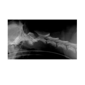
Radiographic Findings
- The radiograph shows a palisade-like periosteal reaction along the vertical body of the atlas.
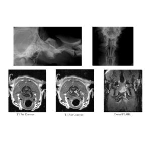
- MRI shows that the suture between the right neural arch and the intercentrum of body of the atlas has ossified normally. The suture between the right and left neural arches has not ossified but the arches retail their normal shape dorsally. The suture between the left neural arch and the intercentrum is wide and irregular with a large buttress of new bone extending ventrolaterally for appox 1.5cm from the left neural arch and a wedge shaped periosteal reaction on the left ventral aspect of the body of the atlas.
- There is no evidence of atlantoaxial subluxation but there is moderate to marked hyperintensity on the STIR and post contrast images in the soft tissue surrounding the lesion including the cleft between the left neural arch and the body of the atlas. There are however, no such changes between the incompletely ossified left and right neural arches themselves dorsally. Importantly, the spinal cord is normal.
Diagnosis
Incomplete ossification of the atlas with surrounding soft tissue inflammation.
This is an uncommonly recognised lesion that has only been recently described in the veterinary literature.
The atlas develops from the fusion of three separate centres of ossification; the right and left neural arches dorsally and on either side and the body ventrally. Failure of the sutures to ossify normally may result in instability between the separate centres of ossification and inflammation of the surrounding tissues may ensue.
Indeed, concurrent anomalies of the surrounding ligaments may predispose to atlantoaxial subluxation. Some dogs may present with neurological deficits referable to C1-C5. Treatment may be conservative or surgical depending on the degree of structural changes present and the severity of clinical signs.
Author
Nikolai Bailey
Morbi leo risus, porta ac consectetur ac, vestibulum at eros. Cras mattis consectetur purus sit amet fermentum. Fusce dapibus, tellus ac cursus commodo, tortor mauris condimentum nibh.