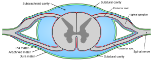
Arachnoid Diverticula
A 4 year 10-month-old male Pug presented with an ataxic gait that had been worsening over the past 3 weeks with subsequent acute onset of dragging the hind limbs, right worse than left. Mild proprioceptive deficits present in both hindlimbs. Spinal reflexes were intact and withdrawal was present. There was no pain on palpation. Forelimbs and cranial nerves all normal. Radiographs taken at the primary care practice showed no significant abnormalities.
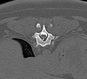
A pre-contrast CT scan of the entire spine did not reveal any significant pathology that might explain the current clinical complaint. No obvious stenosis of the vertebral canal was noted and the intervertebral discs appeared normal and in their correct position.
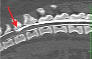
A CT myelogram was then performed by injecting 0.2ml/kg of 300mg/ml warmed Omnipaque into the subarachnoid space at the level of L5-L6. The spine was then imaged in both sternal and dorsal recumbency.
The CT myelogram highlighted a dilation of the dorsal subarachoid space with subsequent stenosis of the vertebral canal at the level of T11-T12 and thus dorsal compression of the spinal cord. This dog was diagnosed with a subarachnoid cyst (also known as an arachnoid diverticulum) at the level of T11-T12.
Surgical decompression of the spinal cord with a dorsal hemilaminectomy and durotomy is recommended for this patient with possible marsupialisation of the diverticulum.
Author
Nikolai Bailey
Morbi leo risus, porta ac consectetur ac, vestibulum at eros. Cras mattis consectetur purus sit amet fermentum. Fusce dapibus, tellus ac cursus commodo, tortor mauris condimentum nibh.